

The world’s smallest automated syringe
The group led by Janos Vörös has developed a nanometer-sized syringe with which medicines, DNA and RNA can be injected into an individual cell without damaging it. In addition to having biological uses, the method could also be applied in the manufacture of microelectronics or microelectromechanical systems (MEMS). The new technology is to be further developed into a marketable product via a start-up company.
Many biologists think the key to life lies in understanding cells. This is because, if we understand how a particular illness originates in cells, and which substances are responsible for which processes, we also know which active compounds should be considered as a therapy. Whereas previously the focus in this search was mainly on cell populations, today it is often on individual cells.
However, the handling of individual cells has hitherto required great manual skill: a potential active molecule is injected into the cell through micropipettes operated using a micromanipulator under a powerful optical microscope. The same apparatus can also be used to measure the tiniest electronic signals in the cell, which in turn allows conclusions to be drawn about the activity of membrane proteins (patch-clamp technique). These proteins are important regulators in cells and play a central role in the efficacy of medicines. Biologists need to be highly experienced, because the method is prone to errors and in many cases the cell is damaged during the manipulation.
A gentle prick thanks to automatic force control
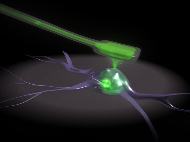
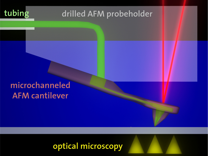
Tomaso Zambelli in the group led by Janos Vörös, Professor at the Institute of Biomedical Technology at ETH Zurich, has presented a nanosyringe for automated injection into cells in the scientific journal “Nano Letters”. Zambelli originally realized that the technology of the atomic force microscope (see box) that is normally used only to image cells could be transformed into a microinjection system. The result was the “fluid force microscope”, according to Zambelli the smallest automated syringe currently in existence. In contrast to a conventional manual system, the pressure exerted on the cell by the measuring needle – also called the cantilever – is adjusted so accurately that the cell is not damaged unnecessarily. A laser is responsible for the control, recording every movement of the cantilever and adjusting the force on the cell several thousand times a second.
This system even operates under water or in other liquids – a precondition for being able to use the instrument to study cells. To enable solutions, too, to be injected into the cell through the needle, scientists at the Swiss Center for Electronics and Microtechnology (CSEM) in Neuchâtel installed a microchannel in the cantilever. The diameter of the opening at the needle tip is only 200 nanometers (approx. 500 times smaller than the diameter of a hair). Substances such as medicinal active ingredients, DNA, and RNA can be injected into a cell through this tip. At the same time, samples can also be taken from a cell through the needle for subsequent analysis.
Observing cells in real time
In an experiment together with Ari Helenius’ group at the Institute of Biochemistry of ETH Zurich, Zambelli has proven that the “fluid force microscope” can be used to inject a virus into an individual cell within a population. They also plan experiments to remove molecules from cells and to measure weak electrical signals at the cell, which in turn allow conclusions to be drawn about the activity of membrane proteins. If the latter should prove possible, biologists would have a practical alternative to the laborious manual patch-clamp technique. In the medium term, the fluid force microscope could be used to enable individual cells to be observed in real time during the injection of active ingredients – and all with the same apparatus. Zambelli explains that, “This would be an enormous advancement for biology and pharmacology research. For the first time, it would enable observing the effect of potential drug candidates on membrane proteins in an automated manner.”
According to Zambelli, this technology has great potential. Well-known manufacturers of atomic force microscopes clearly share this opinion: the scientists have already received offers for the sale of the patent for the fluid force microscope. However, Vörös and Zambelli have decided against this, to allow the two doctoral students Michael Gabi and Pascal Behr in Zambelli’s team, who took part in the development of the fluid force microscope, to continue developing the instrument to marketability in a start-up company founded especially for this purpose, which they called “Cytosurge LLC”. In addition, an initial feasibility study has already been carried out with the help of funding from the Swiss Commission for Technology and Innovation (CTI). Today, Zambelli’s laboratory contains two prototypes of the instrument, which are being tested in collaboration with biologists.
The founders of “Cytosurge” are convinced that there will be a further significant increase in interest in the instrument as soon as it has a greater presence in biological publications. However, in addition to biology, the founders of “Cytosurge” also foresee applications in physics, chemistry and materials sciences. The fluid force microscope opens up new opportunities for the production of increasingly miniaturized microchips in particular, because all kinds of soluble substances and suspensions can be applied to a substrate – and this can even be done in liquids. For example, wafer-thin metallic or electrically conducting polymer tracks can be sprayed onto a surface through the needle tip, thus enabling the construction of nanometer-sized electrical circuits. It would also be possible to etch ultrafine structures out of solid materials using acids as the spray agent.
These ideas still require trials in order to be turned into reality, but the “Fluid Force Microscope” could soon begin a new era for biology in the study of individual cells.
Reference:
Meister et al.: FluidFM:
Combining Atomic Force Microscopy and Nanofluidics in a Universal Liquid
Delivery System for Single Cell Applications and Beyond. Nanoletters. 2009; 9: 2501–2507. DOI: 10.1021/nl901384x
The atomic force Microscope (AFM)
The atomic force microscope is an important tool in surface chemistry and physics. It enables studying the topography of ultrafine structures on a surface using a needle point consisting ideally of only a single atom. This method can be imagined as rather like a record player, in which a needle point traces out the grooves in the disk. The forces acting on the atomic needle and causing the cantilever to bend are measured via a laser beam with which even the tiniest movements of the needle can be recorded and visualized. The atomic force microscope was developed in 1986 by Calvin Quate, Christoph Gerber and Gerd Binnig. In the same year, Binnig and Heinrich Rohrer of IBM Zurich received the Nobel Prize in Physics for inventing the scanning tunneling microscope, which is regarded as the forerunner of the atomic force microscope.
|
|


READER COMMENTS